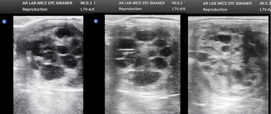
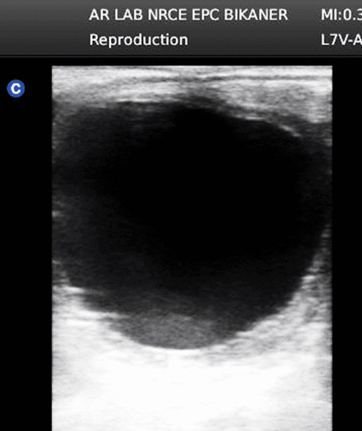
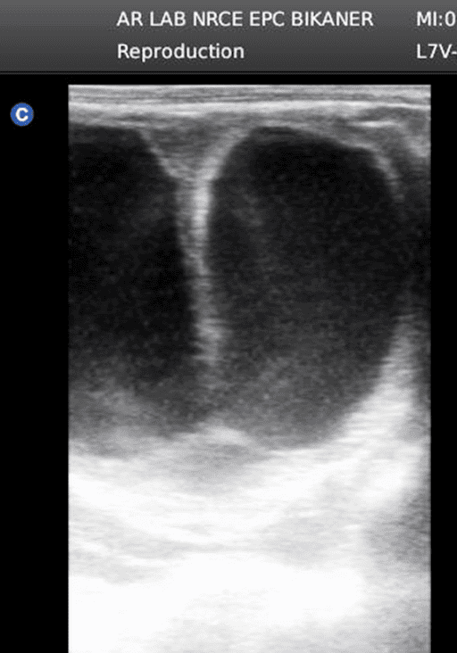
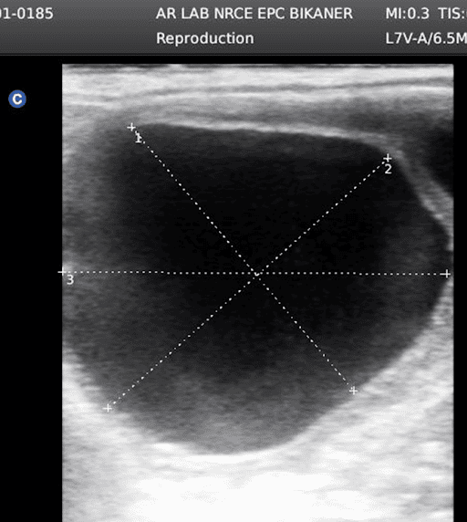
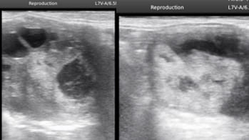
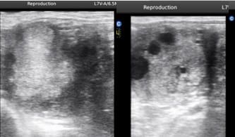
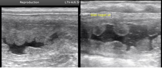
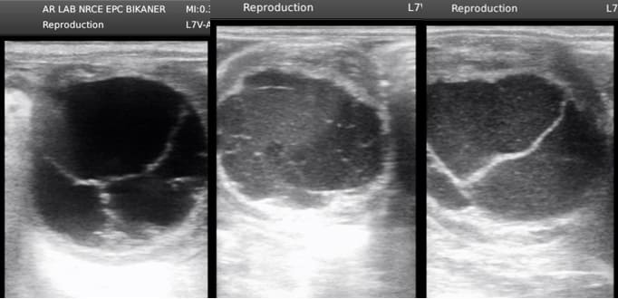
Fig. Pre-ovulatory follicle with specs inside the follicle
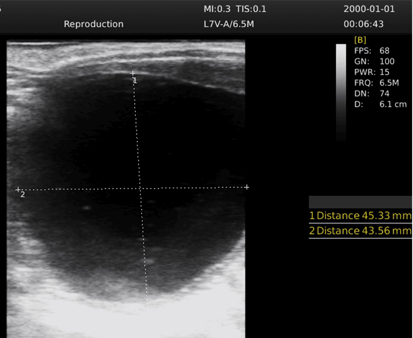
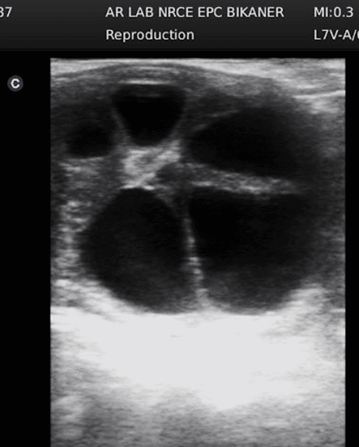
Fig. Uterine/Endometrial Cyst
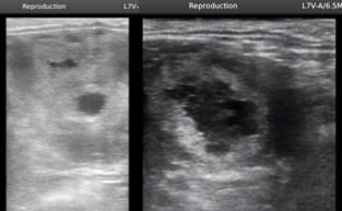
Fig Early pregnancy confirmation through ultrasonography
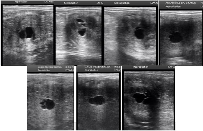
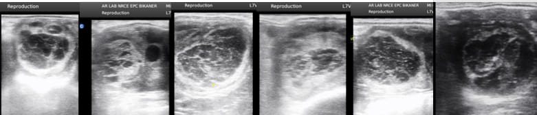
Fig. Anovulatory haemorrhagic follicles on the ovary
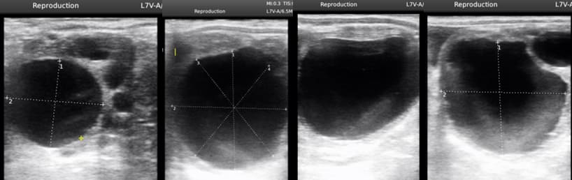
Fig. Pre-ovulatory follicle
Fig. Twin follicles growing together
Fig. Preovulatory follicle indenting towards ovulatory fossa and thickening of the follicular wall near ovulation
Fig. Developing follicles during estrus phase
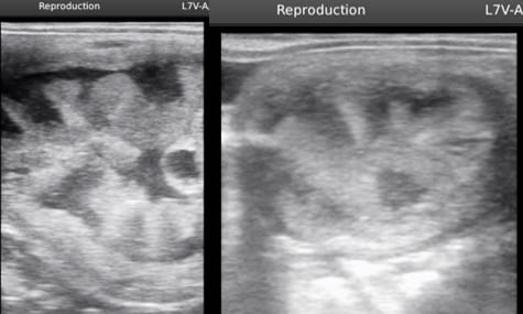
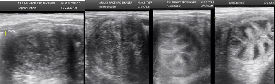
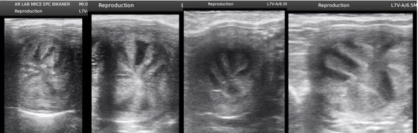
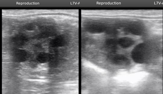
Fig. Uterine edema progressing as estrus phase proceeds
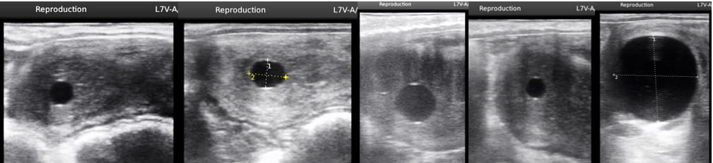
Fig. Progression of the follicle towards ovulation. Note that the spherical follicle becoming irregular near ovulation
Fig. Corpus Luteum
Fig. Double ovulations
Fig. Corpus luteum after imminent ovulation. Notice the fluid within the CL
Fig. Anovulatory follicles during transition period
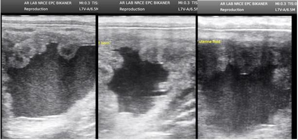
Fig. Accumulation of fluid in the uterus
Fig. Fibrous network development within the growing follicle which will not culminate in ovulation
Fig. Different developmental stages of embryo in mare from day 10 till 18
Fig. Different developmental stages of embryo in mare from day 10 till 30
Fig. Formation of corpus hemorrhagicum (replacement of fluid with blood) after ovulation
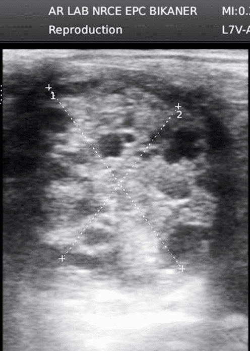
Fig. Typical cart wheel or spoke wheel or cut citrus pattern of uterine folds in mid estrus
Fig. Granulosa cell tumor in ovary
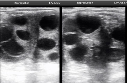
Fig. Inactive ovaries with multiple follicle during winter transition
Fig. Hyperedema observed in some mares during mid estrus. This is normal but sometimes the fluid accumulation persisting and leading to endometritis
Fig. Fluid accumulation in the uterus leading to Uterine infection